Optimizing Image Input
Best practices for image quality, supported formats, and how to capture images efficiently.
Optimizing Image Input
Optimizing your images is crucial for obtaining accurate results with SnapCyte™. Below are the guidelines for capturing high-quality images in the SnapCyte™ App . If you’re planning to upload images to the SnapCyte™ web application, please note that only certain formats are accepted.
Accepted Formats:
- Supported formats include JPEG and PNG.
- Note: TIFF files are not accepted. Please convert TIFF images to one of the supported formats before uploading.
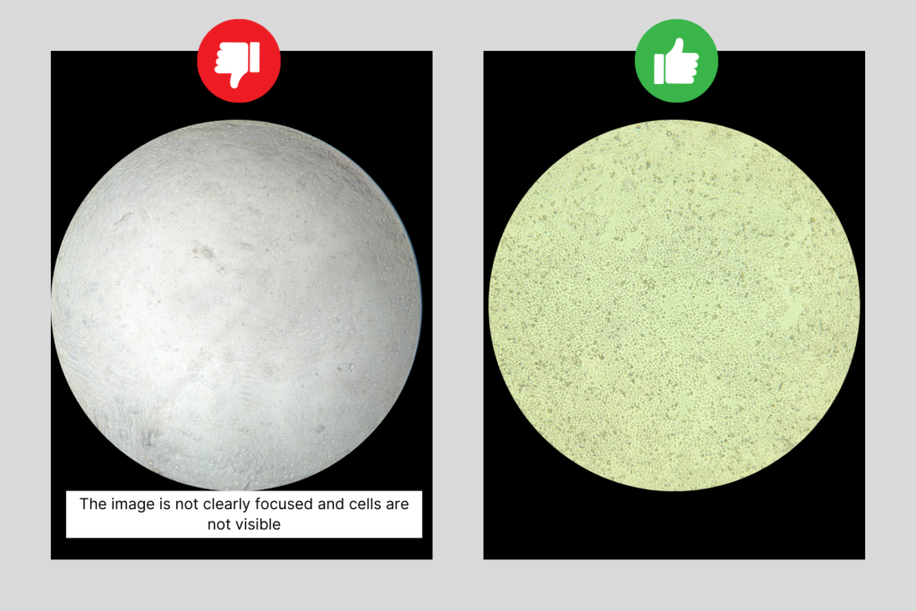
1. Choosing the right magnification and ensuring proper focus
Key Point: The magnification level and focus are both critical to obtaining clear, detailed images of your cells.
Guidelines:
- 4X Magnification: If you can clearly visualize cells at this level, it allows you to cover a larger field of view, capturing more cells in a single image.
- 10X Magnification: Generally recommended as it provides better visualization of individual cells, which can improve the accuracy of your analysis.
- Proper Focus: Always ensure your microscope is focused sharply on the cells. Blurry or out-of-focus images can result in inaccurate analysis, so take the time to adjust the focus until the cells are clear and well-defined.
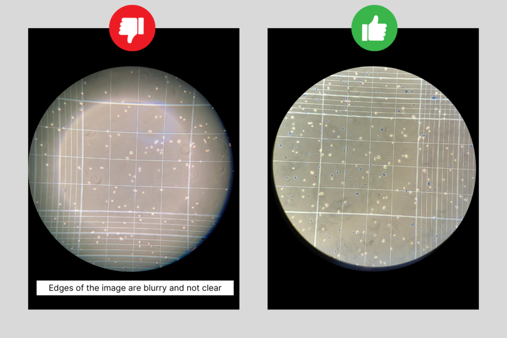
2. Adapter Setup
Key Point: The adapter setup plays a critical role in capturing the best possible image.
Guidelines:
- Ensure that the adapter is properly aligned so that the entire image is visible without any color distortions like blue or yellow rings around the edges. Watch this step by step tutorial on how to set up your adapter.
- Adjust the distance between the phone and the eyepiece to avoid having unfocused rings on the edges.
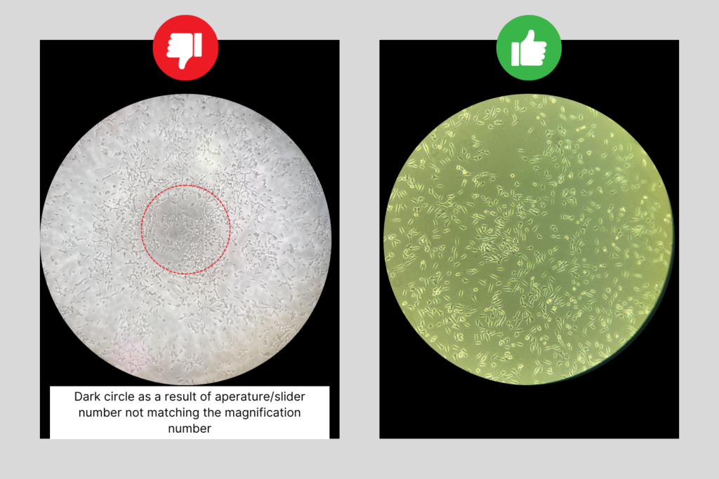
3. Aperture & Slider Adjustment
Key Point: Matching the microscope’s aperture or slider setting with the chosen magnification is essential for achieving the best image quality.
Guidelines:
- Adjust the aperture or slider to control the amount of light passing through your sample, enhancing the contrast and detail visible in the cells.
Video Tutorial: Achieving the Best Image Quality
Watch our comprehensive video tutorial to see these best practices in action.