Our advantage
Cell Proliferation Assay with SnapCyte™
Cell proliferation is crucial for understanding biological processes in cancer research, drug development, and regenerative medicine. Traditional methods like MTT, PrestoBlue, WST-1 and etc, while common, face significant challenges. They require multiple samples to be sacrificed at various time points, leading to cell loss and potential inconsistencies. They are labor-intensive, time-consuming, and can produce variable data due to sample handling.
SnapCyte™ addresses these issues with its image-based, non-invasive technology, to assess cell proliferation with over 95% accuracy. This preserves samples and provides continuous, reliable data.
How SnapCyte™ Works
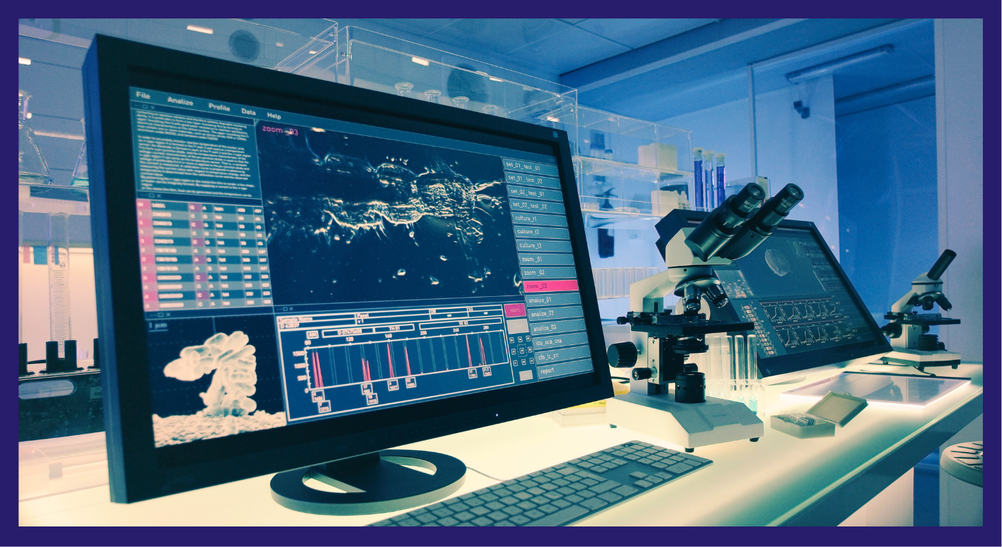
SnapCyte™ streamlines your cell culture analysis process in a few straightforward steps. Start by preparing your samples as usual, culturing and treating your cells to ensure they are ready for imaging at the desired time points. Next, capture or upload high-resolution images of your cell cultures using either your smartphone camera with the SnapCyte™ adapter or a digital microscope. Once the images are uploaded, SnapCyte™’s AI-driven algorithms take over, providing automated and time-lapsed analysis that accurately measures cell density. Finally, review and export the comprehensive data reports generated by SnapCyte™, which include intuitive visualizations and detailed statistical analysis to support your research findings.
Why Use SnapCyte™?
- Save Time and Resources: SnapCyte™ simplifies your workflow, conserving valuable time and resources that would otherwise be spent on traditional assays like MTT, WST-1, XTT, and other similar methods, which require extensive reagent preparation, multiple steps, and longer processing times.
- Enhanced Reproducibility: Ensure consistency in your experiments with AI-driven algorithms that standardize analysis criteria, significantly increasing the reproducibility of your data.
- Comprehensive Record-Keeping: Maintain a thorough and accessible record of your cells, including both images and the analysis, enabling you to track your experiments over time within a single platform.
Request your 30-day free trial
Ready to see the benefits for yourself? Sign up for a free trial and discover how SnapCyte™ can transform your cell culture analysis.
Frequently asked questions
Most Frequent Questions and Answers
What types of cells can I use with SnapCyte™?
SnapCyte™ is compatible with a wide range of adherent cell types making it versatile for various research applications. You can also send sample images for analysis before starting your trial to contact@snapcyte.com.
How many images do I need from each well to obtain accurate, representative data?
The number of images needed per well depends on the format of the culture plate used and homogeneity of your culture. For example, in a 96-well plate, capturing 2 images at 10X magnification per well is typically sufficient for accurate data.
Do I need special equipment to capture images for SnapCyte™?
SnapCyte™ offers an adapter that allows you to acquire images using your smartphone and any basic microscope. It can also work with phase-contrast images taken with other microscopes, providing flexibility in how you capture and upload your data.
Can I track cell proliferation over time with SnapCyte™?
Yes, SnapCyte™ allows for continuous monitoring of cell proliferation by analyzing images captured at multiple time points, providing reliable data throughout your experiments.
How long does it take to capture and analyze images with SnapCyte™?
Analyzing each photo with SnapCyte™ takes less than 30 seconds. For example, if you’re doing an experiment in a 24-well plate, you need to take at least 3 images per well. So, it typically takes about 15 minutes to capture the whole plate.
Is there support available if I encounter issues using SnapCyte™?
Yes, SnapCyte™ offers comprehensive customer support, including detailed protocols, user guides, and direct assistance from our technical team to ensure smooth operation and accurate results.